For the design of Sympfiny, a multi-particulate medicine delivery system for children the company HS Design was subsidized with 50000 $.
Every parent knows that administering medicine to children can be very difficult. The main problem is the awful taste of most medicine. In order to solve the taste problem HS Design offers a high-tech solution with its Sympfiny Design.
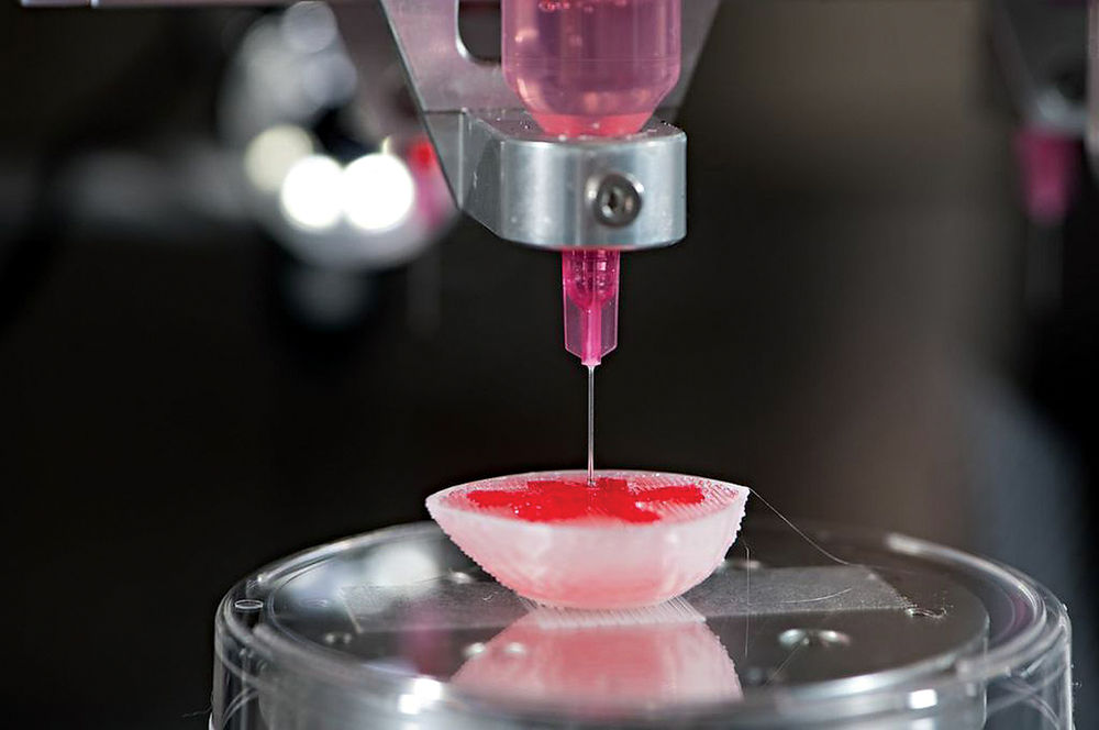
The Sympfiny multi-particulate drug system, delivers medicine not in liquid form but as solid coated microspheres, dissolving in the stomach. In this manner children do not having to deal with the bad taste. The solid drug form offers other problems however. The drug particles can stick together when damp or static, or when being dry some part of the particles can escape through small openings in a pack or satchel. This can lead to inaccurate dose delivery.
Using the same technique for multi-particulate dry powder as for liquid oral medicine, Sympfiny is the new drug-delivery solution. According to Michael Quinn, director of engineering at HS Design, the design of the Sympfiny device is based on the ‘syringe and bottle’ format. The protective bottle stores the multi-particles and the syringe is for dosing.
Solid multi-particulate drugs are also more easy to deliver to children in rural or remote settings in developing countries. The multi-particulate formulation makes medication more easy to store and no water is required for administering the drugs.
After Pfizer and the Institute for Pediatric Innovation worked together in creating the new solid drug formulation technology, it required a different drug delivery system to ensure accurate dosing. The Sympfiny dispensing device was designed ensuring usability, accuracy, and protection of the drug.
At BIOMEDevice Boston, the largest medtech event in New England, the delivery device was selected as one of two winners by Innovation Prize Tour participants. During an open challenge from the Institute of Pediatric Innovation and Pfizer the HS dispensing device design won a $ 50,000 grant.
Autonomic Technologies (ATI), a Californian based company has developed a device for electronic administration of aspirin. With this device patients can administer and control the remote to deliver low-level electrical stimulation for pain relief themselves.
By stimulation of the sphenopalatine ganglion (SPG) nerve cluster the device lingers pain from headache disorders including tension headaches, migraines and cluster headaches. The implant is placed in the upper gum area. With a quite simple procedure the device is inserted causing minimal invasiveness and side effects for the patient.
The permanent implant connects with the SPG bundle of nerves. At the first sign of a headache, the patient can hold the hand-held remote control device on the cheek near the implanted device.
When pressing the remote, the nerve cells are slightly stimulated by an electrical charge blocking the pain signals to the brain. The patient completely controls the device. It can be turned on and off as needed.
Worldwide headache disorders are ranking in third position in causing losing years because of disability. Headache disorders pose an important societal, personal and financial burden. Symptoms such as throbbing pain, dizziness and nausea cause problems in work, family and social life and diminish the quality of life.
The commonly used treatments, such as local anesthetics (xylocaine, lidocaine), analgesics (ibuprofen, aspirin), oxygen therapy, octreotide, phenergan for nausea, sumatriptan and dihydroergotamine intent to lower the severity and number of attacks.
No health restrictions have to be considered with the electronic aspirin and it can be implanted into anyone. The device can be used safely by people with heart disease, high blood pressure or allergies.
According to a clinical study by ATI about electronical aspirin, 68% of patients treated with the device encountered serious improvement. Of these patients 65% experienced no headache disorder anymore within three months.
Electronic aspirin is a renewed technology and still under clinical development, awaiting approval by the FDA. It could possibly become a permanent solution for headache disorders in future.
It is a present-day way of pain relief in society where electronics seem to control a big part of life.
According to a new study, most patients took fewer pain medication after the implantation of a spinal cord Stimulation device. In this study funded by St. Jude Medical their Prodigy Spinal Cord Stimulation System showed that it is effective in treating chronic pain. After receiving the spinal cord stimulation device, the opioid use remained stable or was less than before.
The results lead the researchers to suggest spinal cord stimulation (SCS) to be preferred by physicians over more painkiller prescription for patients whose pain over time got worse. Obstructing pain messages traveling from the nerves to the brain, the small battery-powered transmitters provide signals through electrical wire implanted beside the spinal cord.
The opioid use from 5476 patients with SCS were compared to before and after implantation of the device. One year after implant, the study showed that of the patients continuing SCS therapy, 93% had lower everyday morphine-equivalent doses compared to patients who had their SCS device removed.
Principal researcher and neurosurgeon Ashwini Sharan, stated that patients had an enormous increase in their narcotic use one year before the implant. With the persons continuing with the SCS the dose decreased again afterwards tot the level of before the medication rise.
Unbelievable as it is, the relationship between pain relief narcotics and the implants has never been studied before. The researchers were unaware of which manufacturers’ SCS devices had been implanted in the patients. No funding is provided for further study
According to Sharan, Spinal Cord Stimulation is the last hope because after almost doubling the narcotic dosage within one year, the detachment off this dose takes much lost time.
The cost of a one year morphine prescription is normally $5,000 to which the costs of the side effects have to be added. A spinal cord stimulator averagely cost up to 4 times as much in 2015, depending on the model.
Hospitals tend to choose using the newer models and Sharan says to implant around 300 devices per year, including SCS. According to him he tries to emphasize the distinction between features and function of a device when addressing physicians.
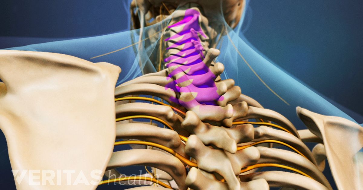
In order to help surgeons perform image-guided open and minimal-invasive spinal surgery, Royal Philips works on an surgical navigation technology based on augmented-reality.
According to a company statement, the technology is significant for cranial and trauma surgeries, pediatric spine surgery, and thoracic spine fusion surgery in adults. Because the thoracic spine is located in the mid- to upper part of the back, the vertebrae are smaller than that of the lower lumbar spine. This makes thoracic fusion surgery more difficult and risky for the surrounding nerves and tissue.
In a preclinical study, more accuracy in placing screws in spines of cadavers, was obtained by neurosurgeons using augmented reality. The accuracy rate of pedicle screw placement in thoracic cadavers’ spines versus freehand placement was increased from 64% to 85% according to the study result.
Less complications are implied by better accuracy according to the business leader of image-guided therapy systems at the company located in Amsterdam.
Since the release of Google Glass in 2013, surgical applications for augmented and virtual reality have gained attention. A London surgeon and the co-inventor of a VR company used the low-cost VR system Google Cardboard to livestream a procedure removing a colon tumor from a patient in April 2016.
In order to add its latest technology, for clinical trials scheduled at about 10 global testing sites, together with its existing low-dose X-ray systems, Philips will need regulatory approval. After full approval by FDA and EU, Philips can expand its use to countless hospitals around the world.
To image the surface of the patient a high-resolution optical camera is placed on a flat X-ray device. The technology constructs a 3D augmented reality vision of the internal and external anatomy, by combining the internal 3D X-ray view and the external camera view. This actual 3D image of the spine of the patient improves procedure planning, surgical tool navigation, accuracy of the implant, and reduces procedure time.
Especially in cranial surgery the technology could be useful, because of brain shape change after releasing the pressure in opening of the cranium. This makes actual images more important than pre-surgical MRI or CT scan images.
Because of the extensive experience in the field of designing image guiding systems Philips needs to tailor them to specific procedures. Starting with spinal surgery, fine tuning the system for trauma surgery will follow, and in the future there is the possibility of the system assisting in brain surgery.
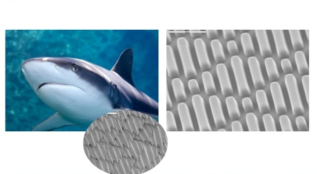
A real innovative surface technology, known as the Sharklet, has been introduced by Sharklet Technolgies Inc. This surface design is inspired by the skin of a shark and reduces bacterial growth without causing resistance, and thus helps protecting for damaging biofilms and microorganisms.
The shark skin technology consists of a series of micro-sized bars interlocking in a diamond pattern. The most important and unique feature of the material is that bacteria accumulation is prevented purely by its surface structure and no chemicals have to be added.
The technology will be mainly used for hospital surface coatings and medical devices.
For which products the Sharklet micropattern design could be beneficial are momentarily being evaluated. Sharklet Technologies has currently partnered up with a medical device firm from China in order to clinically test the micropattern surface technology. This Chinese firm called Peaceful Union wants to advance developing the Sharklet for medical purposes and the surface is now going to clinical trial.
The Sharklet micropattern has shown promising results in clinical pilot studies for further products in the near future.
Sharklet Technologies created the Sharklet in 2007 when a research started about prevention of the coating by algae of marine surfaces, like Naval ships, led to the shark skin pattern. By researching which marine animals skin were not fouling, the unique structure of the sharks skin became apparent.
By experimenting the skin structure was transformed in the Sharklet pattern, It was discovered that no bacterial biofilms were forming on the sharklet pattern and this lead to discovering that no E. coli bacteria were sticking to the pattern and this led to Sharklet Technologies Inc.
The conventional way of killing microorganisms by using disinfectants and antibiotics has led to the creation of resistant bacteria like MRSA for instance. This makes new strategies for managing bacterial growth necessary. By the prevention of bacterial accumulation and growth to begin with, the Sharklet is the perfect solution for this problem.
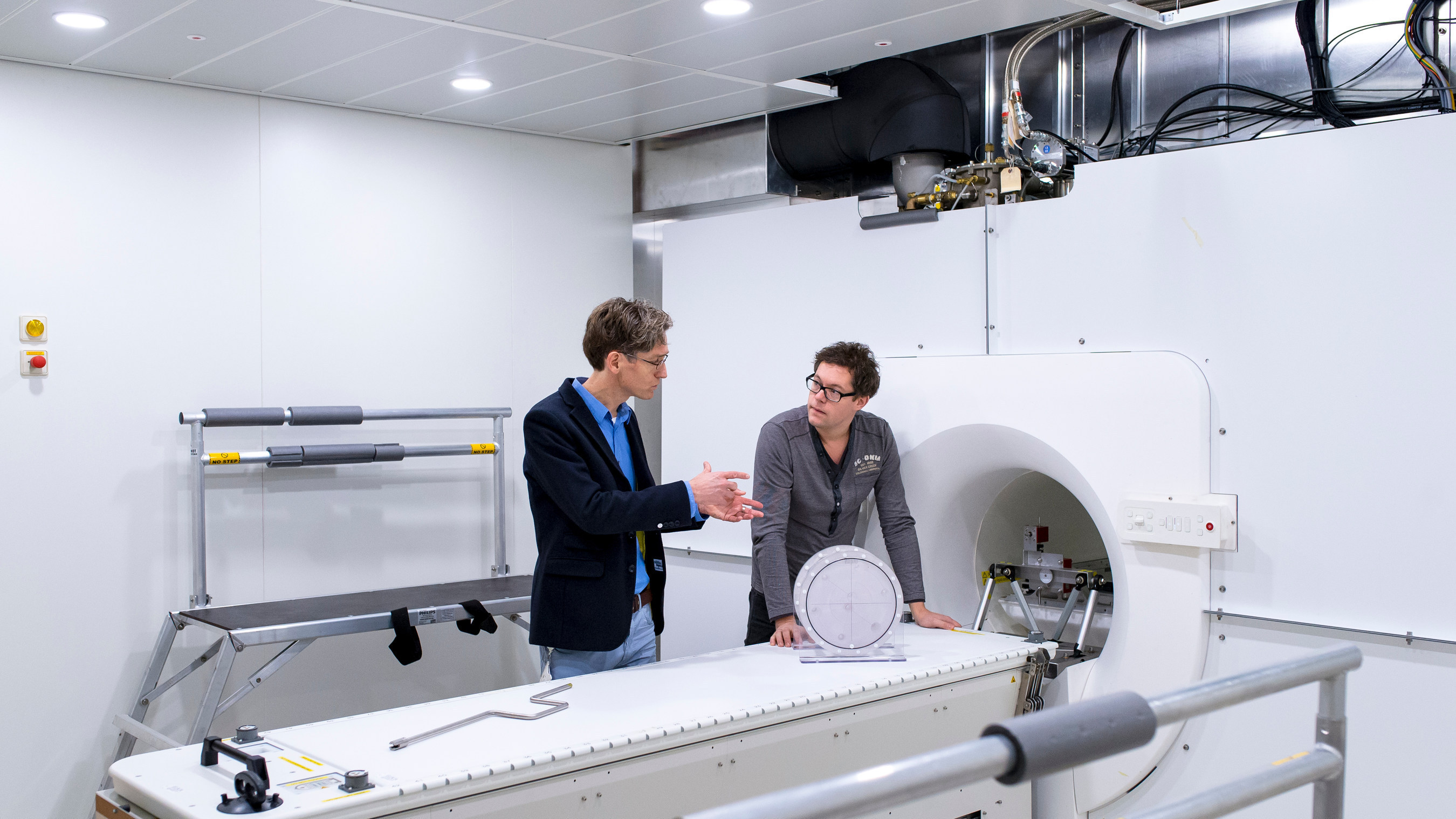
Friday the 19. May the first patient was treated using the high-field MR-guided linear accelerator (MR-Linac) at the University Medical Center (UMC) in Utrecht/Netherlands. This latest radiotherapy device provide accurate and actual visual images enabling precise irradiation with minimal damage to surrounding tissue even in case of a moving tumor while treating.
This MR-Linac system, installed by Elektra and Royal Philips, is emerging as a promising appliance in the field of oncology, for locating and quantifying the tumor tissue, for planning the appropriate therapy, for treatment guidance and therapy evaluation.
According to The Netherlands Cancer Institute, precise and correct dose of radiation is potentially reducing side effects of radiation sessions and improves the quality of life, and ultimately regulating the radiation dose increases tumor control ability.
By combining a MRI device and a radiation device, cancer patients would need less radiation because of the greater effectiveness of the radiation. A further great advantage is that less healthy surrounding tissue is affected by the radiation.
The new device, developed by some Utrecht professors, is currently being tested on people for the first time, after its development period of 18 years. Groundbreaking the experts call this new device and it is expected to be used in hospitals and cancer centers and clinics on a word-wide scale in future. Showing crystal clear images during the treatment, the device can accurately irradiate the tumor, even when moving while the patient is breathing for instance. In this way the treatment can be adjusted to the patient while being in the device. The irradiation times can be lowered so the patient does not have to spend so much time at the hospital.
The experimental irradiation method is currently applied to five patients with bone tumors. When this radiation turns out to be successful, patients with tumors in the abdomen or pelvis are next to receive the treatment.
Digital dentistry which includes state-of-the-art technologies is no longer a fashion, but a necessity.
It offers significant advantages over conventional dental medicine like a more efficient and precise treatment and a much higher predictability level of the final result.
X-rays with a much lower radiation level, a perfectly integrated smile with the face’s features, dental reconstructions performed in record time, even less than 24 hours; these are just a few examples of what modern dental technology can do.
The number of people who have requested complex dental treatments, especially from the field of dental aesthetics has increased in recent years and with demand the supply has also improved. To meet the high demands of the patients, the dental services market has turned to the development of implantology and dental aesthetics through the specialization of doctors and endowment with the latest generation equipment through which they can offer integrated services at international standards.
The aim of a completely computerized way to deal with replicating functional and aesthetic parameters in full arch implant supported restoration procedure is to fully digitalize the entire process.
Digital Technology Benefits
No digital technique is perfect and cannot replace the human hand but the digitalization of dentistry and techniques greatly improves the quality of the prosthetic work greatly fluidizing the steps the patient goes through.
- The tests done are much less as well, as few as two sessions needed to obtain the desired prosthetic work, so the time is reduced to a minimum.
- The processing of prosthetic parts, faster and more accurate, can be done with the help of digital processing equipment and new materials such as zirconium or ceramic type E- Max.
- The finishing of the prosthetic parts is more precise being done by the most advance CADCAM technology. Moreover, nowadays there are special software systems and all digital protocols for dental aesthetics.
- The benefits of digital technologies are felt by the patient from the first session when instead of the classic impression with classic materials hard to bear in the oral cavity he will have a digital scanning.
- Better communication with the patient by being able to visualize their own images and analyze together the contour, symmetries and proportions of the teeth and discuss about possible problems and expectations; in one word, involving the patient in all the steps of the process.
Most Innovative Dentistry Technologies
CADCAM - innovation that enables dental restorations, for example, crowns, veneers, trims and on-lays to be manufactured utilizing electronic processing technology. Dental specialists work with in-office CADCAM to finish same-day tooth rebuilding, efforts that would otherwise require at least two visits to finish. On the other hand, if your case is more complicated, the specialist may work with a dental research facility that uses CADCAM innovation to make the restoration.
There is no cure for type 1 diabetes and many find it challenging to manage this condition. Scientists have come up with a new technology to help better control your type 1diabetes.
Statistics show an alarmingly increased rate of this disease, especially in children.
Type 1 diabetes can fundamentally affect an individual's life, as individuals need to screen their glucose levels consistently to guarantee they are not dangerously high or low.
Individuals with type 1 diabetes measure their glucose levels by pricking a finger a few times each day or wearing a glucose screen. Contingent upon the estimations, they may need to direct insulin utilizing an infusion or insulin pump.
But now scientists are trying a new technology that could replace the traditional methods.
Automatic Insulin System
They targeted a particular kind of artificial pancreas, a closed circle control. These gadgets consistently screen and regulate blood glucose levels. At the point when the screen identifies that an individual needs insulin, a pump discharges the hormone into the body. This trial included the utilization of the Control-IQ system, another kind of fake pancreas that utilizes calculations to modify insulin dosages consequently for the duration of the day.
Specialists needed to replicate everyday life, so they didn't screen the system remotely. Members however contacted researchers at regular intervals to check information from the gadget.
By simplifying the type 1 diabetes management, this innovation could diminish the every day burden of this condition, while likewise conceivably lessening diabetes complications, such as eye and kidney diseases.
Relieving the Burden
The specialists wanted to measure the time that blood glucose levels reached the targeted 70 to 180 milligrams for each deciliter.
The outcomes indicated that the glucose levels of the individuals who utilized the Control-IQ system were in the objective range for about 2.6 hours per day longer than before.
Fundamentally, the system likewise improved the blood glucose control at night just as during the day. This is an essential progression for individuals whose levels drop altogether when sleeping.
More importantly none of the members experienced extreme hypoglycemia when glucose levels become low.
According to researchers these discoveries show that this innovative system can possibly improve the overall health of individuals living with type 1 diabetes, while likewise posibly relieving the burden from those with the disease and their guardians.
The device was just approved for use by the Food and Drug Administration. It will reach patients in January 2020.
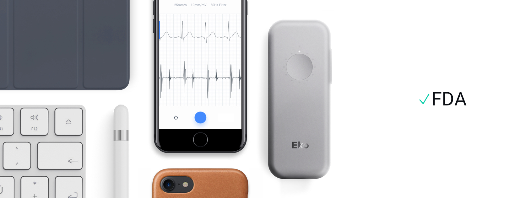
FDA approval was just granted for a new device combining a digital stethoscope and electrocardiogram for home use. This gadget that patients can use at home to automatically alert them and the physicians of aggravated cardiac functions. By developing machine-learning algorithms, in future also a suspected decline in the heart activity is detected.
The handheld device, called the Eko Duo, contains a smartphone app for wireless transmission of heart noise and electrical activity to a specialist as a warning for heart problems.
The stethoscope intensifies heart sounds up to 60 times, contains four digital audio filters and has an enclosed noise reduction. The electrocardiogram (ECG) is to be connected by two electrodes, patients can select between using the 50 Hz or 60 Hz primary filter.
Berkeley’s Eko Duo is a monitoring and warning device for possible heart failure and atrial fibrillation. It detects possible problems, but this doesn’t mean, it could not give a false alarm. According to the company, doctors can use the Eko Duo at the bed or in medical telecommunication consultations with other clinicians.
In developing machine-learning algorithms Eko expects to combine this with Duo to automatically alert care teams and patients of presumed decrease in heart function.
As studies have shown that approximately one quarter of the patients with heart failure are readmitted to hospital within 1 month and half are readmitted within 6 months it’s obvious that cardiac failure cause extremely high costs for the countries.
Cardiologists consider the device as a great way to gather electrocardiographic findings and heart sounds in a user-friendly manner. It may supply cardiologists with data and information from patients concerning the heart sounds that they couldn’t detect by ears in the past.
It is planned to start studies focussed on valvular heart disease, to see if it’s possible to develop an algorithm to reliable recognize of patients with serious valvular disease versus those without. Specialists see a great potential for future development.